Regulation of artery blood pressure
 [PURPOSE]:
Learn how to record the blood pressure (BP) directly.
Observe the effect of neural and humoral factors on BP.
[ANIMAL]:
Rabbit
[METHOD]:
Anaesthesia : 25% urethane solution was injected into the vein in the edge of an ear for anesthesia and the rabbit was fixed on operating table on back.
Separation of common carotid arteries(CCA), vagus never and aortic nerve: The three strips of nerves should be distinguished carefully. The vagus nerve is the thickest, the sympathetic nerve is thinner, and the aortic nerve is the thinnest. The left CCA was used to measure BP and the right nerves were used to be stimulated respectively in the experiment.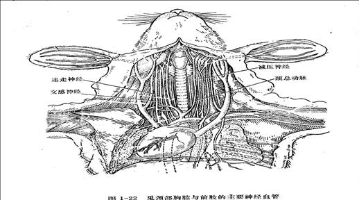
Artery intubatton: Ligate the CCA with thread at the separated segment far from heart, use the arterial clip to clip the segment near the heart. Place a thread under the separated artery with a slipknot for further step. Made an inclined cut toward the heart with an ophthalmologic scissors at the arterial segment 0.5cm distance from the knot, then insert the arterial cannula filled with humoral into the cut towards the heart and fix with the thread prepared, ligate the surplus thread on the side of the cannula to avoid slippage.
Experimental equipments: The output of pressure transducer was connected with the input of computer.
Observation items: Observe the changes of BP when following events happened. Left CCA was cross-clamped for 10 seconds. Aortic nerve was stimulated by a protective electrode.0.3ml 1:10000noradrenalin solution was injected intravenously. Unilateral vagus nerve was ligated and cut off (at the distal end of knot),and the peripheral end of the separated vagus nerve was stimulated.
[Result]:
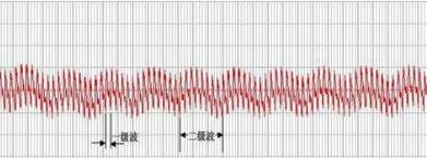
The normal BP
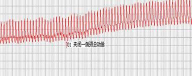
When left CCA was cross-clamped for 10 seconds, according to the pictures, BP is increasing.
When aortic nerve was stimulated by a protective electrode, BP is decreasing.
When 0.3ml 1:10000noradrenalin solution was injected intravenously, BP is increasing.
When unilateral vagus nerve was ligated and cut off and the peripheral end of the separated vagus nerve was stimulated, BP is decreasing. And the curve changes significantly.
[DISCUSSION]:
BP and HOMEOSTASIS
Arterial BP is the pressure that the blood in artery squeezes the vessel. The most important reason why BP forms is that the ventricular contracts and ejects blood to aortic artery.
 Every animal‘s BP is stable relatively. It changed within a certain range. The homeostasis is balanced by neural and humoral regulation. The most important is static artery sinus - aortic arch baroreflex. This reflex can decrease BP. It can also increase BP.As a result, it can reflex BP buffering. The reason why we choose rabbit is that aortic nerve can be separate easily.
So, the first level wave is caused by the fluctuations in ventricular contraction.
The second level wave is caused by the fluctuations in respiratory movement.
LEFT CCA WAS CROSS-CLAMPED
When left CCA was cross-clamped for 10 seconds, according to the pictures, BP is increasing. There is an arterial baroreceptor, in it there are lots of sensory nerve endings. When left CCA was cross-clamped,the carotid sinus baroreceptor receives stimulation to drop . So, the rates of efferent impulses become slower. Then, the nervous of vagus nerve become slower and sympathetic nerve and the vessel of heart are becoming more nervous. These changes make an effect on the heart, so the rate of heart is increasing. The output of heart increase, the vessel of blood contracts and the peripheral resistance of blood vessel increases. At last,BP is increasing.
AORTIC NERVE WAS STIMULATED BY A PROTECTIVE ELECTRODE
When aortic nerve is stimulated by a protective electrode, BP is decreasing.
The aortic nerve of rabbit is afferent nerve.The function of it is that it afferent nerve impulses which is made by aortic arch receptors to medulla oblongata. Then, BP decrease by reflex.
NORADRENALINSOLUTION WAS INJECTED INTRAVENOUSLY
Epinephrine and norepinephrine in the blood are mainly secreted by the adrenal medulla. When vagus nerve is exciting, it can promote adrenal medulla to secrete epinephrine and norepinephrine。
Epinephrine and norepinephrine depends on the role of cardiovascular and vascular smooth muscle cell membrane of myocardial adrenergic receptors on the type and density.
 Adrenergic receptor has two kinds which is α and β. β receptor can be divided into β1 , β2and β3.α receptor excite, and vasoconstriction is the effect. β1 receptors excite and has the effect of "strong heart" . β2 receptors excition makes the vessel diastole. β3 receptors are in adipose tissue.
Epinephrine can activate two receptors α and β, but the effect is stronger on β receptors. β1 receptors in cardiac membrane is effected by epinephrine, and it is the same with cardiac sympathetic nerve. It makes heart beat faster. Conduction speed becomes quicker. Myocardial contraction capacity is more enhancement. Cardiac output increases, and systolic blood pressure increases significantly.
This is due to norepinephrine can combine vascular smooth muscle α and β2 receptors .The blood vessels contract and diameter becomes small,.Peripheral resistance increases. So ,BP increase. In addition, norepinephrine can also increase the heart rate and make cardiac contractility larger. Then, Vasoconstriction is more evident.
UNILATERAL VAGUS NERVE WAS LIGATED
Vagus nerve contain efferent fibers going down from the medulla oblongata , leading to the heart. The end of preganglionic fibers release acetylcholine(Ach) and the end of postganglionic fibers release Ach, too. They belong to parasympathetic fibers. It can reduce the heart rate and atrial contractility decrease. Conductivity decrease.So,the output of heart decrease .As a result, BP decreases.
[CONCLUSION]
The major factors for BP are depressor reflex, effect of cardiac sympathetic and vagus and some medince. Using these can make BP changes. Homeostasis is determined by the neural and humoral regulation.

Laboratory Report

 本组承诺:本实验报告原创,绝无抄袭!

第二篇:机能实验学实验报告书写格式示例20xx0423-护理学
机能实验学实验报告书写格式示例
日 期:20##年12月5日
20##年春季学期 机能实验学实验报告
日 期: