生物系实验报告

(Prohibited for commercial purposes)
姓名 班级 学号 实验日期 科目 实验名称 the cell cycle and mitosis 合作者 指导教师 成绩
LAB 6: The Cell Cycle and Mitosis
Introduction:
Mitosis is the main process of cellular reproduction. It contains interphase, metaphase, anaphase and telophase; cytokinesis included. Two identical daughter cells which nearly possess the same genetic materials in parent cell. Interphase is a phase including G1, S, and G2, which is not part of mitosis. The chromosomal materials whose DNA have been fixated can be dyed by Feulgen stain. According to the root tips cells we observe, the different mitotic stages can be easily distinguished.
In this lab, we can observe different stages of mitosis in plants cells. We will make a wet mount slide of garlic root tips, so that we can learn to use selective staining techniques (only stain DNA in chromosomes)- Feulgen stain.
Materials and methods:
Materials: Compound microscope (NIKON ECLIPSE E100), water bath (at 60℃), glacial acetic acid-methanol fixative (1:3), 1M hydrochloric acid, 45% acetic acid, Feulgen stain, prepared slides of onion root tip, lens tissue, garlic root tips, forceps, slides, cover slips.
Methods:
Part Ⅰ: ①Observe a prepared slide of a longitudinal section of onion root tip with microscope, from
low-power to high-power.
②Concentrate in the region where the apical meristem of the root, and identify cells in different
phases.
③Look at 30-50 onion root tip cells, then tally the frequency of mitotic phases. Record the data
in table T-1.
Part Ⅱ: In order to prepare a wet mount slide.
.①Add 4 ml of freshly prepared methanol-acetic fixative in a vial, then deal with about 2-3 mm garlic root tips in it for 15 minutes at 60℃.
.②Having finished fixative, slowly pour off the fixative in to waster container, then add 2 ml 1M hydrochloric acid to the vial.
.③Place the vial in the water bath for 10 minutes at 60℃, so that the plant cell walls can be

.④Pour off the acid, add 1ml Feulgen stain into the vial, maintain for 45-60 minutes at room temperature.
.⑤Place a drop of acetic acid on the slide, and transfer a root tip (cut from the root of garlic) to it, then cover the sample with coverslip.
.⑥Dry the slide with two pieces of paper towels, use thumb to press directly downward over the coverslip. Be careful to prevent the cells from rolling and overlapping.
.⑦Observe the slide with low power to locate the pink region of chromosomes which is stained. Reduce the light intensity to see the details of cells and chromosomes.
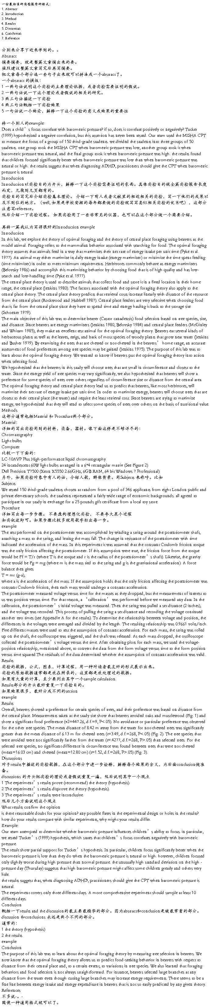
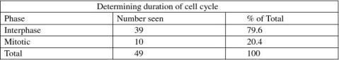
.⑧Study the slide and identify the cells in different mitotic phases. Look at 30-50 dividing garlic root tip cells and calculate the frequency of the phases, then record the data in table T-2. Results:
PartⅠ: As we can see in a series of Figures 1-1 and table T-1, some regions of root are composed of normal
cells and the area that contains many cells of mitotic phase is the apical meristem. 79.6% of cells are in interphase while there are 20.4% of cells in mitotic phase, and overview the cells which are in telophase; we can see the unclear cell plate. Cell plate can finally grow to cell wall which completely separates the two daughter cells.
PartⅡ: According to Figure 2-1, 2-2, and table T-2, the cells which are in mitotic phase (metaphase,
anaphase, telophase included) occupy 19.2%. When the cell is in metaphase, its chromosomes will arrange on the equator of it. But the chromosomes tend to move to the two poles of cells and we can see the separation clearly. As telophase, we can see cells temporarily with two nuclei. On the basis of what we know, the nucleus of a cell in prophase starts to disperse, but it is hard to recognize whether or not. So the proportion of the cells in prophase is 70.8%, similar to the interphase in PartⅠ,much larger than reality. At the same time, the data shows that the percentage of metaphase, anaphase and telophase is 6.2%, 9.2%, and 13.8% respectively.
Discussions:
1. What is the function of mitosis in cell cycle?
Mitosis is nuclear division plus cytokinesis, it allows a cell inherit its traits to the two daughter cells which have nearly same chromosomal materials as it. Because it ensures the equal division of chromosomes, and keeps the stability of the organism.
2. Besides Feulgen stain, please suggest other methods to visualize chromosomes in mitotic cells. As what we know, other DNA stain like methylrosaniline chloride (gentian violet), acetone-carmine, and methyl chloride can also dye DNA, with different of similar mechanism. Besides, maybe we can also directly use high-power electron microscope.


References:
Baidupedia, lab manual.
Distribution statements:
HE Weiwei is responsible for controlling the time and record the phenomena what we see, and LIU Zhichao make the slide of garlic root tip. At the same time, Lin Jirong is the one who observe the slide with light microscope and take photos with hiss windows phone.
Figures:
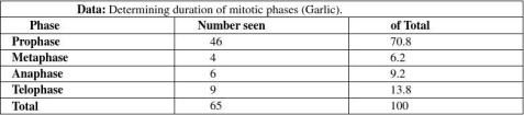
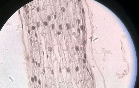
A B
Figure 1-1.1 (10×40)
The data in table T-1 is based on 3 columns of cells in this figure. A with 2 nuclei represents the cell in mitotic phase, and B represents the cells in interphase.
Figure 1-1.2 (10×40) Figure 1-1.3 (10×40)
They are the cells in different region of onion root tip.

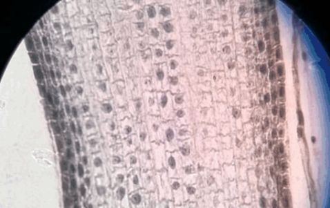
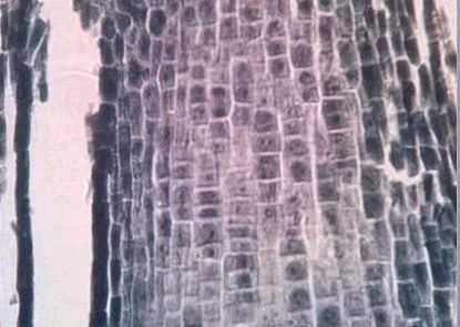
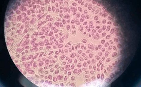
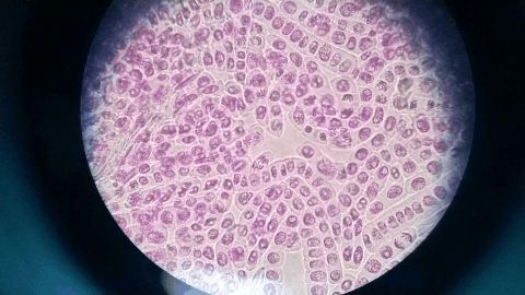
Figure 1-1.4 (10×40) Figure 1-1.5 (10×40) These pictures are taken at different location of the onion root tip.
B
Figure 2-1 (10×40)
A is the cell in metaphase, and B represents the cell which is in anaphase.
Figure 2-2 the whole view.


第 页
第二篇:英文实验报告的格式和写法
英文实验报告的格式和写法【转】
20##-10-04 06:03